Syphilis, as one of the sexually transmitted diseases, brings immense suffering to patients, not only causing psychological pressure but also leaving them at a loss, too afraid to go to the hospital for a check-up. However, if this disease is left untreated, it will progress to secondary syphilis. So, how is secondary syphilis diagnosed and examined?

Diagnostic Criteria for Secondary Syphilis:
1. Medical History: A history of extramarital sexual contact or infection from a spouse. There may be a history of primary syphilis, which generally occurs 6 weeks to 6 months after infection or 6 to 8 weeks after the appearance of a chancre.
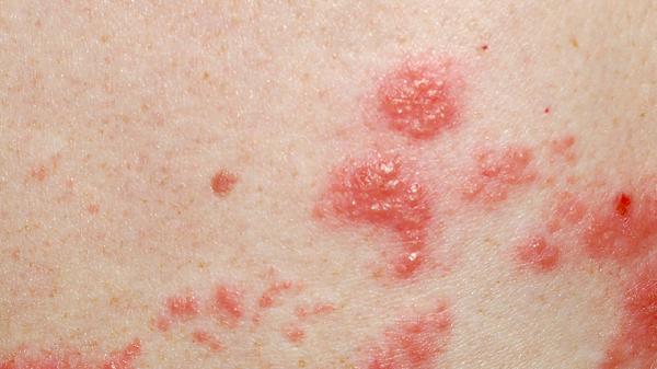
2. Various skin rashes such as roseola, papules, flat condylomas, palmoplantar syphilids, mucous patches, and moth-eaten alopecia, accompanied by general discomfort and lymphadenopathy.
3. Dark-field Microscopy: Treponema pallidum can be detected in the exudate of flat condylomas, moist papules, and mucous patches.
4. Syphilis Serological Tests: Tests such as RPR, TPHA, or FTA-ABS are strongly positive.
Differential Diagnosis:
Condyloma Acuminata: Condyloma acuminata is a sexually transmitted disease, over 95% of which is caused by unsafe sexual intercourse. Its incubation period is 2-3 months, and its appearance is cauliflower-like, mushroom-like, or cockscomb-shaped, slightly sharper and harder than pseudocondylomas, and more fragile, easily bleeding upon contact. If an acetic acid test is performed, the papules will turn white.
Vitiligo: The skin lesions are depigmented patches, often milky white or light pink, with smooth surfaces and no rashes. The borders of the white patches are clear, with increased pigmentation at the edges compared to normal skin. The hair within the white patches is normal or turns white.
Drug Eruption: The skin rash is not obvious, resembling large-area burns, with scaling. The skin appears healthy, but when gently rubbed with a finger, the epidermis peels off in large areas.
Additionally, it should be differentiated from diseases such as psoriasis, lichen planus, folliculitis, impetigo, and thrush.
Laboratory Examinations for Secondary Syphilis:
1. Dark-field Microscopy: Treponema pallidum is easily detected in secondary rashes, especially flat condylomas, moist papules, and mucous patches. Tissue exudate or lymph node puncture fluid is scraped with a slide to observe active Treponema pallidum.
2. Syphilis Serological Tests (non-treponemal antigen tests and treponemal antigen tests) are strongly positive.
3. Biopsy for Treponema pallidum, using silver staining (Warthin-Starry) or fluorescence antibody staining, can reveal Treponema pallidum, appearing dark brown with a spiral structure, located around dermal capillaries.
4. The Unheated Serum Reagin (USR) test is also a modification of the VDRL antigen, with sensitivity and specificity similar to VDRL.